The Inefficiency of Oral Supplementation
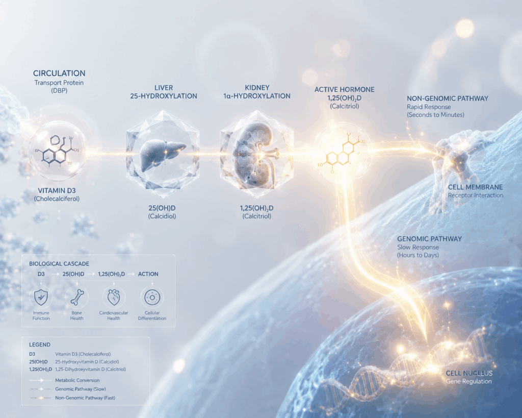
Ingesting a standard D3 capsule does not yield immediate physiological effects. The human body requires a strictly regulated two-step hydroxylation process to activate the molecule. Without optimal function at both stages, the prohormone remains biologically inert.
Hepatic Hydroxylation: Primary Processing
Whether synthesized cutaneously or absorbed enterically, D3 initially travels to the liver. Hepatocytes utilize the enzyme 25-hydroxylase (CYP2R1) to attach a hydroxyl group (–OH), converting D3 into 25-hydroxyvitamin D [25(OH)D]. Because this initial step is not rate-limiting, the liver processes the total available supply of raw D3.
Once hydroxylated, 25(OH)D enters systemic circulation with a half-life of two to three weeks, maintaining relatively stable concentrations. This is the precise molecule measured during clinical laboratory testing, as it provides the most accurate reflection of the body’s total reserve.
Renal Activation: Final Synthesis
Circulating 25(OH)D lacks intrinsic biological activity. It requires subsequent hydroxylation within the kidneys, governed by the enzyme 1α-hydroxylase (CYP27B1) under the strict regulation of parathyroid hormone (PTH). This reaction yields 1,25-dihydroxyvitamin D [1,25(OH)₂D], also known as calcitriol, which is the biologically active hormone.
Calcitriol binds to VDRs globally to initiate gene transcription, directly managing calcium kinetics, cellular differentiation, and neurotransmitter production. Because its half-life spans merely four to six hours, it acts as an acute biological signal. Clinicians generally avoid measuring 1,25(OH)₂D for baseline assessments because it reflects transient physiological demand rather than sustained nutritional status.
Endogenous Regulatory Mechanisms
Elevated concentrations of 1,25(OH)₂D naturally induce the expression of 24-hydroxylase (CYP24A1) in target tissues. This distinct enzyme degrades excess 25(OH)D and 1,25(OH)₂D into inactive, excretable metabolites. This continuous degradation pathway prevents clinical toxicity even when daily intakes reach 5,000 IU.
Substrate Availability Constraints
Macrophage defensive capacity is mathematically limited by systemic 25(OH)D concentrations. At a serum level of 15 ng/mL, activated macrophages simply lack the raw substrate required to synthesize adequate 1,25(OH)₂D, rendering TLR distress signals ineffective. In such cases, susceptibility to infection stems from an acute substrate deficit rather than an inherent immune defect.
Epidemiological data consistently show that infection rates for respiratory syncytial virus, influenza, and COVID-19 surge during winter months, mirroring the seasonal nadir in population-wide vitamin D levels.
Dual-Channel Signaling Kinetics
In the classical genomic pathway, 1,25(OH)₂D enters the nucleus, binds to the VDR, forms a heterodimer with RXR, and targets specific VDRE sequences on DNA to alter gene transcription. This cascade typically requires hours or days to manifest observable physiological changes.
Recent molecular analyses confirm that a subset of VDRs localize directly to the cell membrane and cytoplasm. Binding at these sites triggers non-genomic cascades within milliseconds. This facilitates the rapid release of calcium ions, the activation of protein kinases, and the opening of trans-membrane ion channels, providing a critical mechanism for acute cellular stress responses.
D3 vs. D2: Structural Variances and Efficacy
The commercial market frequently offers Vitamin D2 (ergocalciferol) as a cost-effective, plant-and-fungi-derived alternative to D3.
Parameter | D3 (Cholecalciferol) | D2 (Ergocalciferol) |
Primary Source | Endogenous human synthesis / zoological | Botanical / fungal |
Serum 25(OH)D Elevation Efficiency | High | Diminished by 30–50% |
Pharmacokinetic Half-life | Extended | Truncated |
Impact on All-Cause Mortality | Validated by Cochrane meta-analysis | No substantive evidence |
Clinical Recommendation | Yes | No |
Given the distinct pharmacokinetic disadvantages of ergocalciferol, clinical protocols strongly favor cholecalciferol exclusively.