
Two-Step Unlocking: How D3 Actually Works in the Body
And Why Most Supplements Only Make It Halfway
Vitamin D is rarely considered beyond its presence on a calcium supplement label or a routine physical exam report. However, modern urban lifestyles have contributed to a widespread, yet largely ignored, systemic insufficiency. In 2019, Jikei University School of Medicine in Tokyo screened 5,518 healthy adults using the gold-standard LC-MS/MS method. The screening revealed that 98% of the subjects maintained serum vitamin D levels below 30 ng/mL, highlighting a critical gap in urban health management.
The Origins of a Century-Old Error
American biochemist Elmer Verner McCollum discovered a fat-soluble substance in cod liver oil in 1922 capable of curing rickets. He designated it “Vitamin D.” This nomenclature was subsequently adopted across medical literature and diagnostic frameworks, despite being fundamentally inaccurate.
A true vitamin is defined by two rigid criteria: the human body must be unable to synthesize it endogenously, and it must be acquired through diet. Vitamin D3 meets neither requirement. The human epidermis stores 7-dehydrocholesterol. Upon exposure to UVB radiation (wavelengths of 290–315 nm), a sunlight-driven steroid synthesis pathway is initiated automatically. This specific mechanism has been conserved in the genetic architecture of vertebrates for over 500 million years, predating the need for dietary supplementation entirely.
Biological Classification
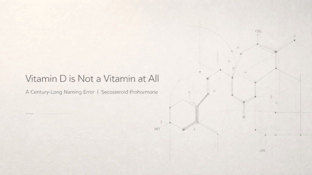
The precise biochemical classification of vitamin D3 (cholecalciferol) is a seco-steroid prohormone.
The term “seco-steroid” accurately describes a molecular structure featuring an open B-ring, distinguishing it from closed-ring classic steroid hormones like estrogen, testosterone, and cortisol. Despite this structural variance, its metabolic pathways, receptor binding affinities, and signal transduction profiles are functionally identical to traditional hormones. It operates not as a basic nutrient, but as an advanced signaling molecule regulating a network spanning more than 200 distinct cell types.
Systemic Receptor Distribution
The vitamin D receptor (VDR) is present in nearly every nucleated cell in the human body. As a member of the nuclear receptor superfamily—alongside thyroid and retinoic acid receptors—the active form of D3 systematically integrates into broader gene expression networks.
VDR Localization | Primary Regulatory Function |
Cerebral cortex, hippocampus, substantia nigra | Memory retention, mood stability, motivation, neurogenesis |
Cardiomyocytes, vascular smooth muscle, endothelial cells | Blood pressure homeostasis, vascular integrity, cardiac output |
T cells, B cells, macrophages, dendritic cells | Immune activation thresholds and suppression timing |
Pancreatic β cells | Insulin secretion dynamics, blood glucose regulation |
Ovaries, testes, placenta | Sex hormone synthesis, reproductive fertility |
Osteoblasts, osteoclasts | Skeletal mineralization and bone turnover |
This extensive receptor distribution underscores its physiological importance far beyond conventional skeletal health.
Navigating the Literature
Insufficient serum vitamin D levels compromise multiple physiological systems simultaneously. To understand the full scope of this molecule, the subsequent sections of this series will detail the following clinical aspects:
- The rigorous two-step endogenous hydroxylation required for physiological activation.
- The mechanism by which immune cells locally synthesize active D3 to combat pathogens.
- The diagnostic superiority of specific blood testing methodologies over generalized advice.
- The critical, yet frequently overlooked, role of essential cofactors such as K2 and magnesium.
- A standardized protocol for assessment, dosing, and long-term clinical monitoring.